Accuracy and Personalized Care
At Catholic Health, we know that early detection of heart disease is critical to accurate diagnosis and customized treatment plans. Cardiac imaging is offered at all Catholic Health hospitals across Long Island.
Cardiac imaging can diagnose a range of heart conditions. We use the most advanced technology to detect a heart condition early, diagnose with more accuracy and guide the most effective treatments.
Board-certified cardiologists supervise and interpret all procedures. In addition, our caring staff guides you through every aspect of your imaging test, so you know what to expect before, during and after your test. We work with your referring physician to create a tailored imaging plan and, in most cases, we provide results to your physician on the same day.
Advanced Diagnostic Imaging
St. Francis Hospital & Heart Center (Roslyn, NY) is a leader in cardiovascular diagnostic imaging. The hospital's team of cardiac experts makes it possible to image the heart with such great detail and speed that a diagnosis can be made in seconds without an invasive procedure.
You can also find the same level of care at the St. Francis Heart Center at Good Samaritan University Hospital (West Islip, NY).
Appointments
St. Francis Hospital & Heart Center (Roslyn, NY)
- For Cardiac Nuclear Imaging and Echocardiography (Echo): 516-705-6627
- For CT and MRI: 516-563-7100
Good Samaritan University Hospital (West Islip, NY)
- Call 631-376-4440
Imaging Services
Not all services are offered at all locations.
- Cardiac amyloid
- Cardiac iron overload
- Cardiac sarcoid
- Cardiac tumor
- Cardiomyopathy
- Congenital heart disease
- Congestive heart failure
- Coronary artery disease
- Mitral valve disease
- Pericardium disease
Cardiac Magnetic Resonance Imaging (MRI)
Advanced MRI technologies offer high-speed and high-resolution images of the beating heart. These images accurately illustrate the heart chambers, heart muscle, wall motion, ejection fraction and blood flow. Cardiac MRI is used to diagnose a wide range of heart conditions, including coronary artery disease, congenital heart defects and other problems of the heart muscle and pericardium.
A Cardiac MRI is noninvasive, does not use radiation and takes about 45 minutes. With some exceptions, it’s safe for those with cardiac stents, artificial valves and metal implants. Plus, our skilled and compassionate team is experienced at helping reduce claustrophobic anxiety.
Cardiac Computerized Tomography (CT)
Advanced CT instantly provides 3D, motion-free images of the beating heart. The images captured detail the coronary artery anatomy, including bypass grafts, plaque features and stenosis severity. A computerized tomographic angiography (CTA) combines CT technology with an injection of a contrast dye to create detailed images of the blood vessels.
These images allow our physicians to diagnose and treat disease more accurately. High-speed imaging also minimizes your breath-hold time.
Our team also provides expertise in:
- Complex vascular imaging
- Congenital heart disease
- Left- and right-ventricular function
- Pre- and post-TAVR evaluation
- Pre- and post-Watchman device implantation assessment
- Pulmonary vein mapping
Cardiac Nuclear Imaging
Nuclear imaging is helpful for those who are not candidates for simple stress tests, which involve exercise. The addition of nuclear imaging increases the diagnostic accuracy of the stress test, especially when coronary artery disease is suspected.
Single-photon emission computed tomography (SPECT) is a specialized type of nuclear imaging. It involves an injection of nuclear isotopes and continuous imaging by a gamma camera that circles the body. This approach may help identify coronary artery disease in women, who often experience false positives with stress tests, as well as older patients. The information from this scan can help our cardiologists determine who is a candidate for coronary angiography, angioplasty and stenting, or coronary artery bypass surgery.
Echocardiography (ECHO)
An echo uses ultrasound, or sound waves, to create pictures of the heart. These pictures are more detailed than standard X-ray images. Specialized testing includes:
- Transthoracic echo—most common type of echo
- Interventional echo—guides valvular and other percutaneous procedures
- Intraoperative echo—assists our surgical team with surgery planning
- Transesophageal echo—uses a probe inserted into the mouth and down the esophagus
- Stress echo—combines ultrasound with a stress test
All applicable studies include rigorous, specialized 3D analyses to ensure accuracy in assessing myocardial and valvular function, blood flow, hemodynamics and anatomical investigation.
Coronary Calcium Score
A quick and noninvasive scan of your coronary arteries can save your life.
Cardiac Imaging Locations
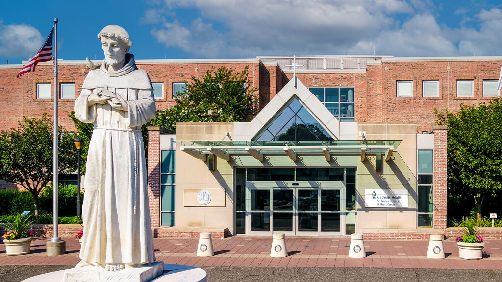
St. Francis Hospital & Heart Center
Roslyn, NY
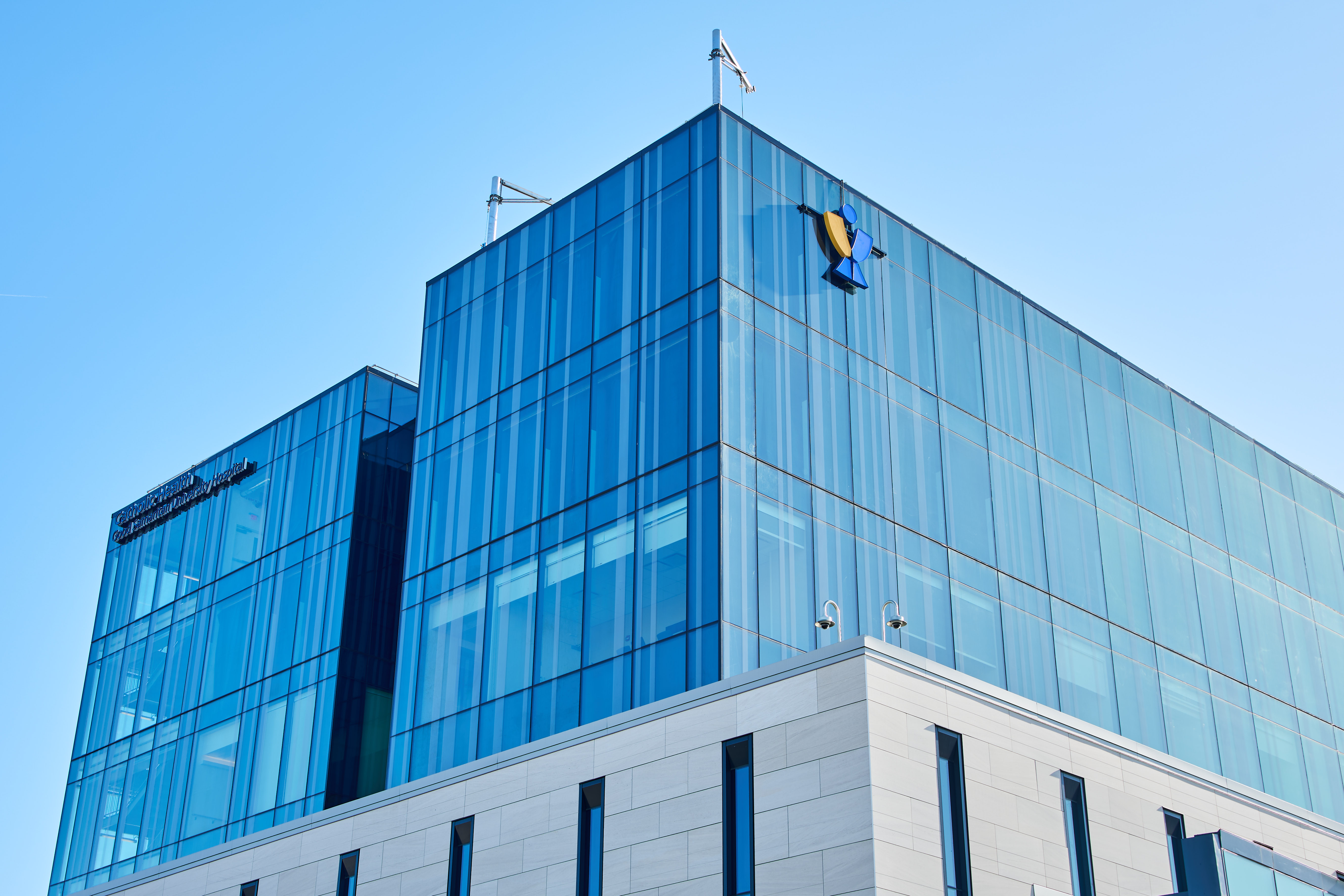
St. Francis Heart Center at Good Samaritan University Hospital
West Islip, NY
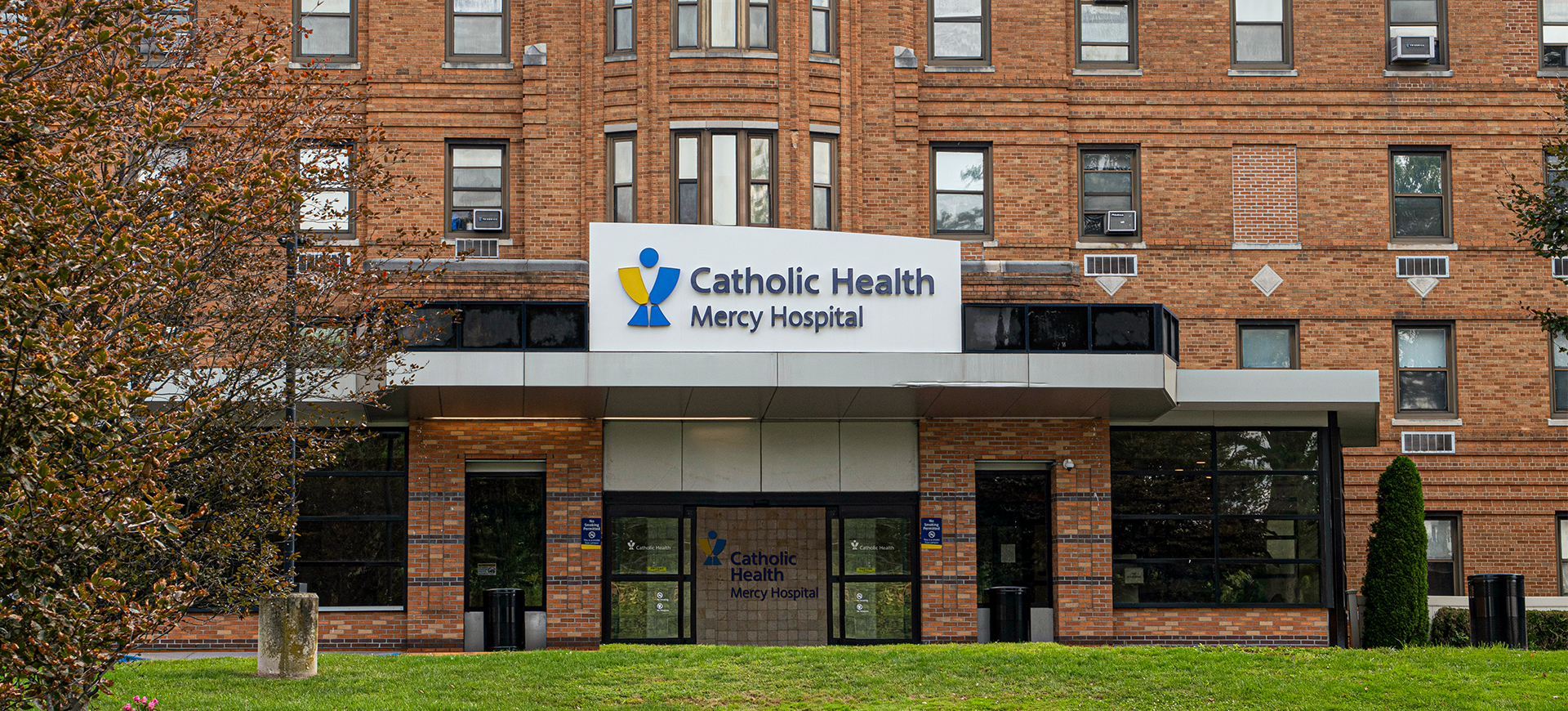
St. Francis Heart Center at Mercy Hospital
Rockville Centre, NY
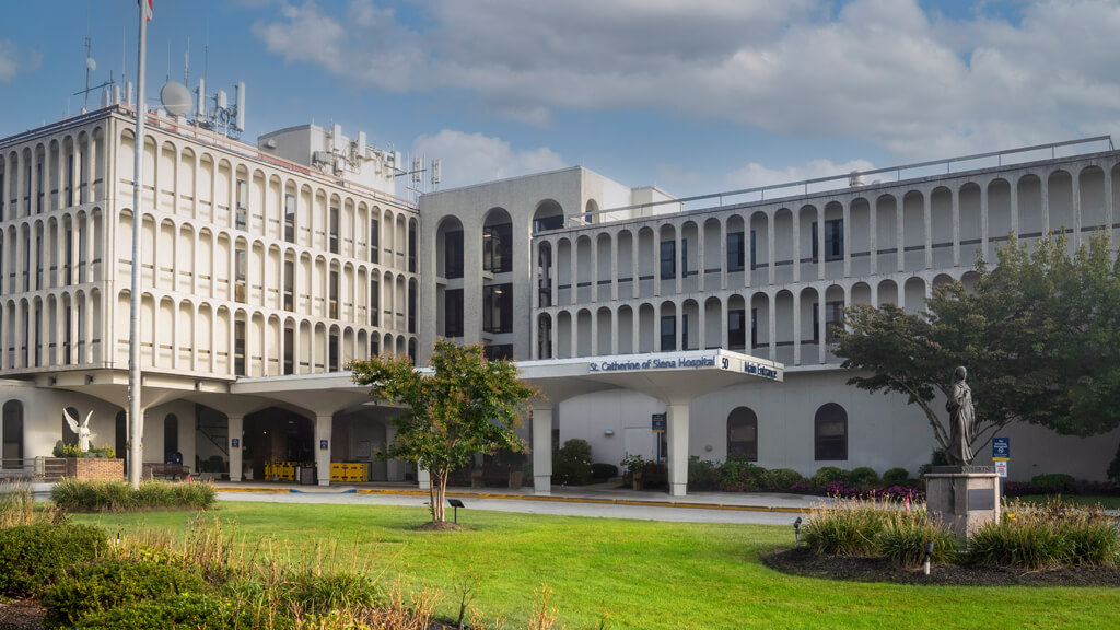
St. Catherine of Siena Hospital
Smithtown, NY
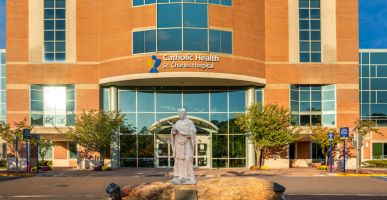
St. Charles Hospital
Port Jefferson, NY

